Dear Geremia,
Here is the procedure I use for S4L:
Prerequisites:
The Yale Neuron compiler 8.2.6 for Windows
https://github.com/neuronsimulator/nrn/releases/tag/8.2.6
You can use newer versions, but this one works best with S4L.
Create a directory for a particular neuron structure, at root level: hoc files, asc, etc. a subdirectory called 'mechanisms': all your mod files.
From a console, run the Yale Compiler on your 'mechanisms' directory:
> nrnivmodl
if it succeeds, it will create a nrnmech.dll file.
move the nrnmech.dll file to root level (where your hoc files are) and clean the 'mechanisms' directory - all the .c and .o files.
Compress into a zip file the main directory (that contains the hoc files, nrnmech.dll and 'mechanisms' dir). Change the extension to .hocz
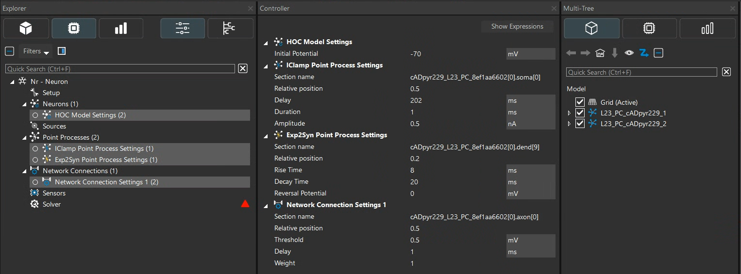
Now, in S4L use the import button to import your new neuron model, your newly created hocz file.
Regards,
Guillermo